Collaterals
Children who are born with complex congenital heart disease associated with a reduced blood flow to their lungs can sometimes develop collateral vessels.
What are collaterals?
If a baby is born with a malformation of the heart and a lack of blood flowing to the lungs to collect oxygen, the child will have low oxygen saturations (the amount of oxygen in their blood). They are cyanosed (have blue-coloured lips and fingernails) and may be breathless on mild exercise or feeding. Collaterals are connections, like normal blood vessels, that can develop in children with cyanotic heart disease such as single ventricle heart conditions.
There are two types of collaterals:
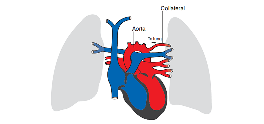
Systemic Arterial Collaterals
These abnormal vessels originate from the body blood vessels, in particular the Aorta, and grow towards the lungs. They can form when a child has had a long period of cyanosis (blue lips and fingernails due to low oxygen levels in the blood). The collaterals aim to take more blood to the lungs where it can collect oxygen. This is the body’s response to the long standing low oxygen saturations. These collaterals make the child less blue but create more work for the heart!

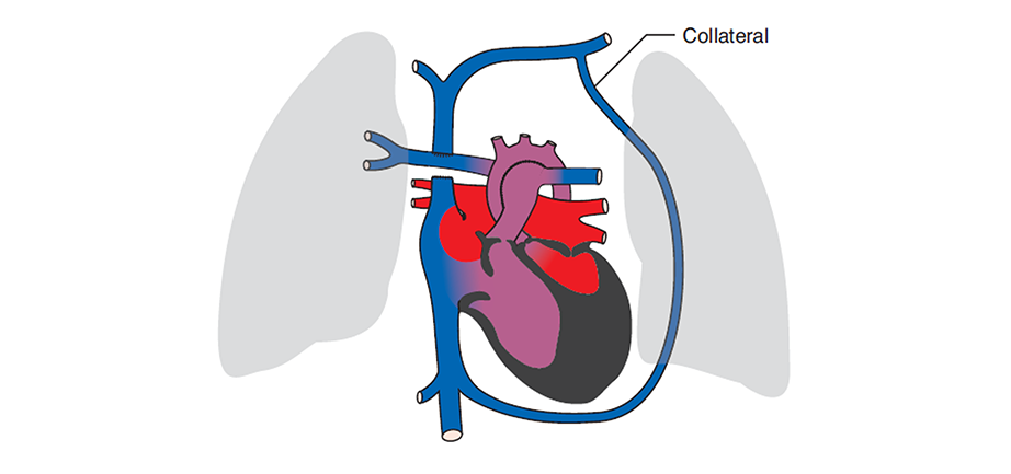
Systemic Venous Collaterals
These are abnormal blood vessels that originate from the veins taking the blue (deoxygenated) blood back to the heart. They normally develop after the second operation, the Cavo Pulmonary Connection (or Stage Two). After this operation the pressure in the veins in the upper body is greater than the pressure in the veins in the lower body. With that, small veins can enlarge and can allow blue blood from the upper body to run down to the lower body rather than having to squeeze through the lungs. These collaterals make the child more blue, but do not increase the work for the heart.

How are collaterals diagnosed?
When children with single ventricle disorders undergo cardiac catheterisation or MRI scanning investigations it is possible to clearly see the collateral vessels. When there is a large collateral vessel it may be seen during a routine echo (echocardiogram). See Cardiac Tests.
How are collaterals treated?
Once collateral vessels have been found the cardiac team will assess if they need closing (occlusion). Small systemic arterial collaterals will normally disappear after the Fontan procedure has been performed. Large systemic arterial collaterals should normally be closed by a catheter procedure as they put strain on the heart and raise the pressure in the lung arteries. The child may be more cyanosed after this, and the Fontan operation may have to be performed earlier.
Large systemic venous collaterals in young children after the Cavo Pulmonary Connection operation (Stage Two) should be closed by catheter. This will make them less cyanosed, as more blue blood goes to the lungs, and frequently the Fontan operation can be delayed. Small systemic venous collaterals identified just before the Fontan operation can be ignored.
Page uploaded September 2023